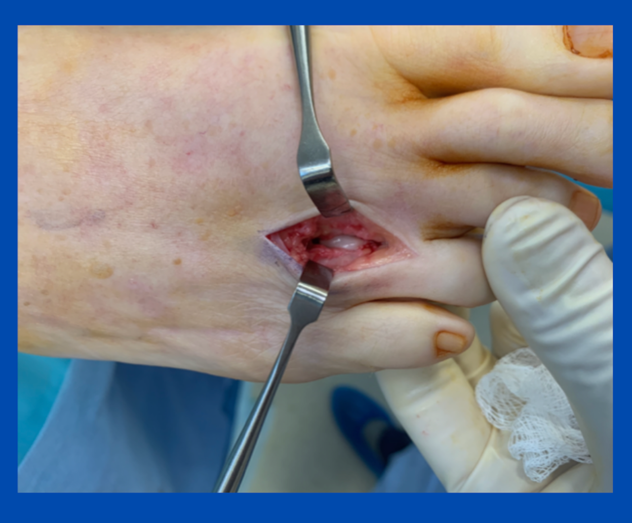
Neuromas are one of the most common conditions seen by a podiatric surgeon. The image is of a recent case, showing how small the incision can be and what a neuroma looks like intra-op. The shiny white structure in the wound is the neuroma I have pushed up with my left index finger to deliver it into the wound.
One of the main advances in neuroma surgery is the release of the deep transverse ligament. The neuroma sits deep to this and is not intermetatarsally as animated images often show. The neuroma also sits slightly distal to the metatarsal heads. I have found this knowledge and subtle tweak in the surgery allow us to achieve a higher success rate and marked reduction in stump neuroma occurrence.
This also allows a shorter recuperation process. However, it is still imperative that a large compression bandage is applied, and the patient carries out minimal weight bearing the first two weeks. Suture knots are removed at this stage, and the patient is typically out of the postoperative shoe at approximately three weeks.
If you have any specific questions or would like to discuss similar cases, feel free to contact me.
(This content is intended for healthcare professionals only)
