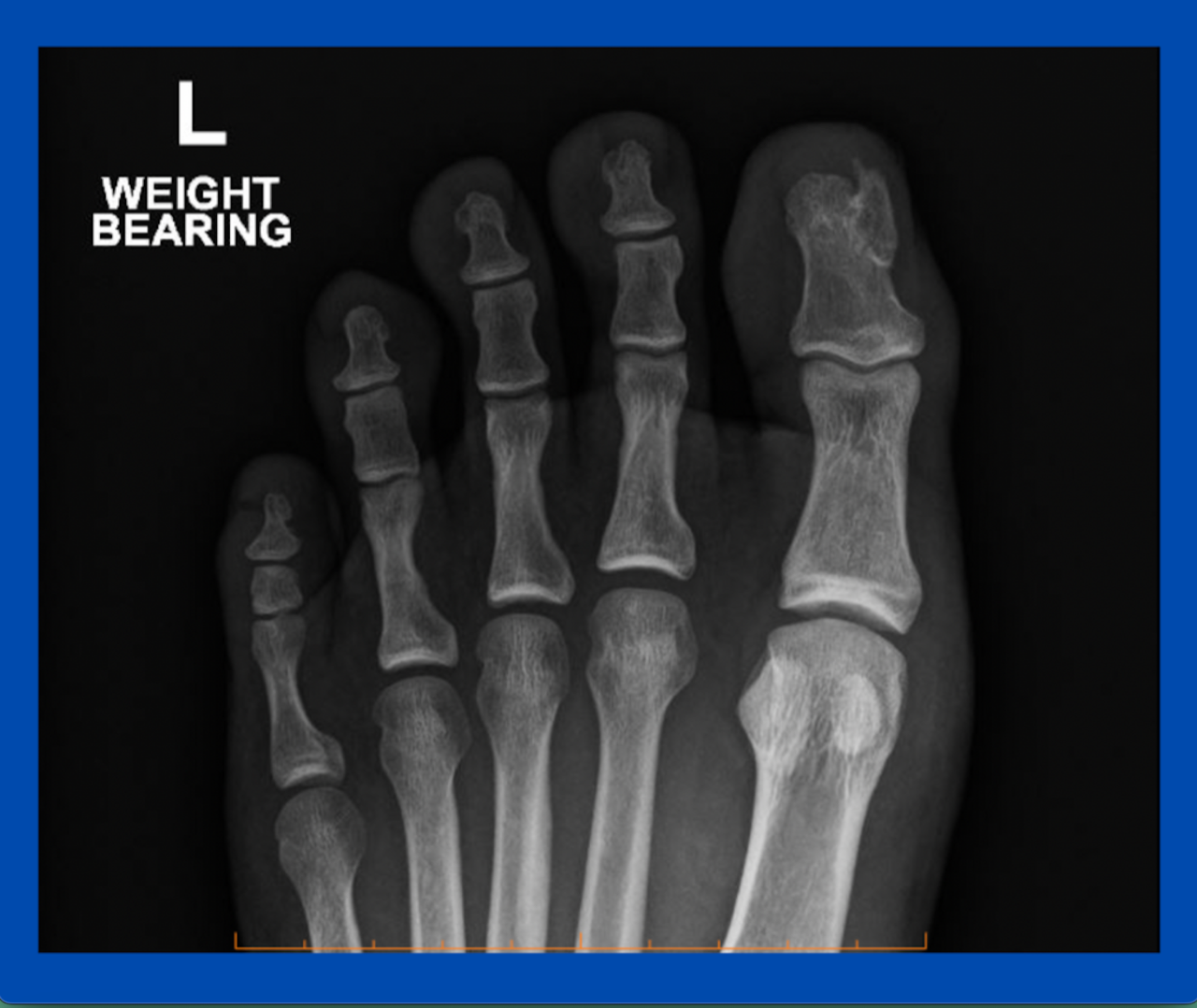
This is one of a few subungual exostoses I see regularly throughout the year. Often when I see these cases, they have been treated as an ingrown toenail for some time.
I find they have a very particular appearance (a firm fibrous/callus on top) and sit just under the nail.
Sometimes they can present on the sulci and can impinge on the nail leading to hypergranulation. This, I think, could be the reason they are often mistaken for ingrown toenails.
Subungual exostoses are nearly always osteochondromas that are benign (noncancerous) bone tumours. In fact, they are the most common bone tumour. They have a bony base with a cartilaginous cap and tend to continue to grow until they pierce the skin, resulting in marked pain and infection, as bacteria are given a portal to enter the body and infect the surrounding tissue.
Subungual exostoses are easily diagnosed with a plain X-ray as can be seen in the image. Surgery is typically advised as they tend to continue to grow. The procedure is quite straight forward involving local anaesthetic with a small incision just distal the prominence. As long as a small amount of normal bone is removed just deep to the bony pedicle, I find they rarely return. The recovery is quick and all pain should resolve.
If you have any specific questions or would like to discuss similar cases, feel free to contact me.
Also read:
Hypergranulation tissue
Remember functional anatomy and to x-ray
(This content is intended for healthcare professionals only)
