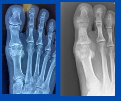
This patient presented approximately two weeks ago with a history of long-term gout. He is only in his thirties, and his history of supposed gout flare-ups does not seem like your typical gout presentation.
He has marked symptoms; however, the 1st MTPJ is never red, hot and swollen. He had blood tests many years ago, which showed evidence of hyperuricemia. This has hence been labelled as a gouty joint with treatment revolving around this diagnosis, with drugs being the mainstay. This has not resolved his pain, and the joint ache and stiffness have progressed.
A thorough history, physical exam and radiographic assessment were carried out at my office. All the above leads me to believe this patient has been wrongly diagnosed with gout. The imaging below shows his imaging on the left. The right is an example of a radiograph with gout. Note the signs of erosions and shadowing of tophaceous deposits in the joint with chronic gout. I suspect he has degenerative osteoarthrosis/hallux limitus.
Further investigation could involve aspiration of the joint to assess for urate crystal deposition; however, I am sure he has hallux limitus, which has a very different treatment regime to gout.
If you have any specific questions or would like to discuss similar cases, feel free to contact me.
Also read:
Gout deposits (not bunions)
Gout
Remember functional anatomy and to x-ray
(This content is intended for healthcare professionals only)

