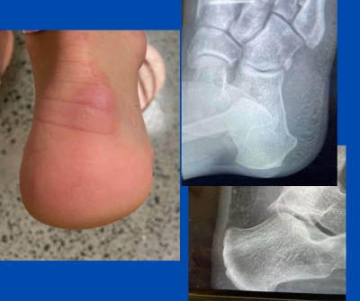
This patient presented with a history of markedly painful posterior heel “bumps”. She has noted them for approximately two years. The patient is now having major problems with any enclosed shoes. The area rubs become red and enlarged and can blister. She feels she has exhausted conservative care. She is young and is keen to return to enclosed shoes.
Various causes exist, including insertional tendonosis, Haglund’s deformity and retrocalcaneal exostosis.
X-ray and ultrasound show, luckily for the patient, there is no achilles tendon involvement, as can be seen by the imaging below. She has small osseous protrusions on the lateral posterior calcaneus.
As it is NOT deep in the achilles tendon, minimally invasive exostectomies would allow the bumps to be burred off through small incisions, with very quick recuperation. If the lumps involved spurring in the achilles insertion or it was a true Haglund’s, the surgery would be much more involved and require a very long recovery process, including initial non-weightbearing in a backslab for six weeks.
This is another example of medical imaging allowing for accurate diagnosis.
If you have any specific questions or would like to discuss similar cases, feel free to contact me.
Also read:
Ultrasound in the operating theatre
Anterior Ankle Impingement Syndrome
Remember functional anatomy and to x-ray
(This content is intended for healthcare professionals only)

