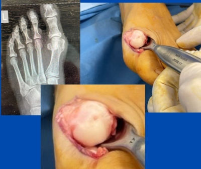
I operated on this patient last week. She had suffered from marked left 1st MTPJ pain.
She had been treated for her bunion via rest, orthotics, mobilisation, medication and various splints. No pain relief was achieved, and she noted that the joint was becoming slightly stiff and more symptomatic.
X-ray assessment showed a bunion (hallux abducto valgus); however, it was only moderate, and symptoms were more marked than expected with a bunion of this size. On examination, the patient winced on motion and guarded the joint.
As the symptoms were marked and the joint was becoming stiff, I suspected there was cartilage damage from excess and abnormal joint motion. The patient does, however, remember trauma to the joint. The patient felt she had exhausted non-surgical options and was keen to have her “bunions corrected”.
An MRI could have been prescribed to identify synovitis, capsulitis or osteochondral defects, but this would be an extra cost for the patient. I was sure there was cartilage damage, plus the patient was keen for surgery; therefore, an MRI was not carried out.
As can be seen, by the imaging on the right and the close-up, there was indeed an osteochondral defect. Once this was “cleaned up”, the area of damaged cartilage was approximately twice as big as the initial image below depicts. During the surgery, the bunion was corrected, the defect debrided, and the subchondral bone fenestrated to allow cells the migrate into the defect. Once the bunion deformity was corrected, the cartilage damage dealt with, and the wound closed, platelet-rich plasma (PRP) was injected into the joint to help stimulate fibrocartilage to grow into the defect.
If you have any specific questions or would like to discuss similar cases, feel free to contact me.
Also read:
Hallux Limitus / Rigidus
Post-operative wounds
Dislocated 2nd MTPJ
Platelet Rich Plasma (PRP)
(This content is intended for healthcare professionals only)

